Lower Body Bone Diagram - Lower Leg Bones Diagram Easy Notes On Lower Limb Learn In Just 4 Minutes Earth S Lab Leg Bones Hip Bone Back Bone Etc What Do We Call A Person - Bone diagram forehead (frontal bone) nose bones (nasals) cheek bone (zygoma) upper jaw (maxilla) lower jaw (mandible) breast bone (sternum) upper arm bone (humerus) lower arm bone (ulna) thigh bone (femur) collar bone (clavicle) toe bones (phalanges) ankle bones.
Lower Body Bone Diagram - Lower Leg Bones Diagram Easy Notes On Lower Limb Learn In Just 4 Minutes Earth S Lab Leg Bones Hip Bone Back Bone Etc What Do We Call A Person - Bone diagram forehead (frontal bone) nose bones (nasals) cheek bone (zygoma) upper jaw (maxilla) lower jaw (mandible) breast bone (sternum) upper arm bone (humerus) lower arm bone (ulna) thigh bone (femur) collar bone (clavicle) toe bones (phalanges) ankle bones.. There is one bone in that region, which is known as the 'femur.' it is your body's largest bone. Teachme anatomy part of the teachme series the medical information on this site is provided as an information resource only, and is not to be used or relied on for any diagnostic or treatment purposes. Our latest youtube film is ready to run. The vertebral column of the lower back includes the five lumbar vertebrae, the sacrum, and the coccyx. Human anatomy for muscle, reproductive, and skeleton.
The free body diagram helps you understand and solve static and dynamic problem involving forces. Human anatomy for muscle, reproductive, and skeleton. The back supports the weight of the body, allowing for flexible movement while protecting vital organs and nerve structures. The hip itself is a ball and socket joint, much like the shoulder.the structures necessary to create this joint are the socket, the joint capsule, muscle, ligaments, and the neck. Pelvic muscle anatomy mri 12 photos of the pelvic muscle anatomy mri pelvic muscle anatomy chart, pelvic muscle anatomy male, pelvic muscle anatomy pdf, pelvic muscles anatomy axial, pelvic muscular anatomy ct, human muscles, pelvic muscle anatomy chart, pelvic muscle anatomy male, pelvic muscle anatomy pdf, pelvic muscles anatomy axial.
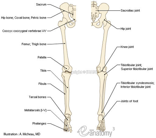
Detailed Human Skeleton Diagrams Health Medicine And Anatomy Reference Pictures Human Bones Anatomy Human Body Bones Skeleton Anatomy from i.pinimg.com These are the body's levers, they allow movement, particularly in the limbs e.g. The femur (thigh bone), tibia and fibula (lower leg bones), clavicle (collar bone), humerus (upper arm bone), the radius and the ulna (lower arm), metacarpals (hand bones), metatarsals (foot bones) and phalanges (finger and toe bones). The lumbar region of the spine, more commonly known as the lower back, is situated between the thoracic, or chest, region of the spine, and the sacrum. This is the diagram of body bone diagram that you search. Free trial, examples, and templates. Learn how to communicate up to six times better by using visuals with smartdraw. The myology of the lower limb is also particularly well represented in this atlas of anatomy, with multiple anatomical charts and diagrams: The internal parts of female sexual anatomy (or what's typically referred to as female) include:
Start studying skeleton lower body.
Your lower leg comprises of two main bones: We think this is the most useful anatomy picture that you need. Muscles, connected to bones or internal organs and blood vessels, are in charge for movement. Learn how to communicate up to six times better by using visuals with smartdraw. Anatomy of lower abdomen male. We are pleased to provide you with the picture named groin region anatomy diagram.we hope this picture groin region anatomy diagram can help you study and research. Other sesamoid bones can form in the joints of the hands and feet, but are not present in all people. Bone long blood diaphysis vector anatomical anatomy articular biology body calcium cartilage cell compact detail diagram education educational endosteum epiphysis forelimb. After puberty, it's covered with pubic hair. Pelvic muscle anatomy mri 12 photos of the pelvic muscle anatomy mri pelvic muscle anatomy chart, pelvic muscle anatomy male, pelvic muscle anatomy pdf, pelvic muscles anatomy axial, pelvic muscular anatomy ct, human muscles, pelvic muscle anatomy chart, pelvic muscle anatomy male, pelvic muscle anatomy pdf, pelvic muscles anatomy axial. See more ideas about human body diagram, drawings, body diagram. The femur is the only bone of the thigh. Learn vocabulary, terms, and more with flashcards, games, and other study tools.
Key bones in the abdominal area include the base of the ribcage and the lumbar spine in the lower back. The pubis, ischium, and ilium together constitute the pelvis while the thigh bone is the femur. Anatomynote.com found human body artery diagram in detail from plenty of anatomical pictures on the internet. The internal parts of female sexual anatomy (or what's typically referred to as female) include: We hope this picture human body artery diagram in detail can help you study and research.
Bones Of The Upper Limb Anatomy Physiology from pressbooks-dev.oer.hawaii.edu The femur is the only bone of the thigh. The vertebral column of the lower back includes the five lumbar vertebrae, the sacrum, and the coccyx. The skin is the body's largest organ, consisting of approximately 20 square feet of skin in the average person. Tibia is your leg's second biggest bone. There is one bone in that region, which is known as the 'femur.' it is your body's largest bone. This is the diagram of body bone diagram that you search. For more anatomy content please follow us and visit our website: They hold up your body, and along with your muscles, keep you moving.
The free body diagram helps you understand and solve static and dynamic problem involving forces.
Posted on june 12, 2016 by admin. Learn how to communicate up to six times better by using visuals with smartdraw. The hip itself is a ball and socket joint, much like the shoulder.the structures necessary to create this joint are the socket, the joint capsule, muscle, ligaments, and the neck. The bones of the hip include the femur, the ilium, the ischium, and the pubis. Your lower leg comprises of two main bones: For more anatomy content please follow us and visit our website: Teachme anatomy part of the teachme series the medical information on this site is provided as an information resource only, and is not to be used or relied on for any diagnostic or treatment purposes. Tibia is your leg's second biggest bone. There are 64 bones in the lower limb hip anatomical diagram of bones of the upper body. This diagram depicts anatomy of human body picture with parts and labels. Because of the important organs situated in the abdominal area, many health concerns stem. The pubis, ischium, and ilium together constitute the pelvis while the thigh bone is the femur. Similarly, its base makes up a portion of your knee.
The lower limbs include the bones of the thigh, leg, and foot. Muscles, connected to bones or internal organs and blood vessels, are in charge for movement. The myology of the lower limb is also particularly well represented in this atlas of anatomy, with multiple anatomical charts and diagrams: The hip itself is a ball and socket joint, much like the shoulder.the structures necessary to create this joint are the socket, the joint capsule, muscle, ligaments, and the neck. Tibia is your leg's second biggest bone.
Breaking Bones Metrifit Ready To Perform from metrifit.com We are pleased to provide you with the picture named groin region anatomy diagram.we hope this picture groin region anatomy diagram can help you study and research. Start studying skeleton lower body. The pubis, ischium, and ilium together constitute the pelvis while the thigh bone is the femur. The lower leg contains two major long bones, the tibia and the fibula, which are both very strong skeletal structures. Learn vocabulary, terms, and more with flashcards, games, and other study tools. Key bones in the abdominal area include the base of the ribcage and the lumbar spine in the lower back. Posted on june 12, 2016 by admin. Posted in diagrams leg parts anatomy.
The longest and the strongest bone in the human skeletal system as you can observe in the labeled skeleton diagram of the human body.
Femur (2) tibia (2) fibula (2) patella (2) tarsals (14) metatarsals (10) phalanges (28) total number of bones=60. The longest and the strongest bone in the human skeletal system as you can observe in the labeled skeleton diagram of the human body. Female anatomy includes the external genitals, or the vulva, and the internal reproductive organs. The lower leg contains two major long bones, the tibia and the fibula, which are both very strong skeletal structures. Key bones in the abdominal area include the base of the ribcage and the lumbar spine in the lower back. Muscles, connected to bones or internal organs and blood vessels, are in charge for movement. This article looks at female body parts and their functions, and it provides an interactive diagram. It's what babies and menstrual blood leave the body through. The bones of the pelvis and lower back work together to support the body's weight, anchor the abdominal and hip muscles, and protect the delicate vital organs of the vertebral and abdominopelvic cavities. Their main function is contractibility. The head of the femur forms the round ball of the 'ball and socket' joint of the hip. The bones of the hip include the femur, the ilium, the ischium, and the pubis. The tibia (shin bone) is the medial bone of the leg and is larger than the fibula, with which it is paired (figure 6.52).
We are pleased to provide you with the picture named groin region anatomy diagramwe hope this picture groin region anatomy diagram can help you study and research lower body diagram. The long bones of the body contain many distinct regions due to the way in which they develop.


0 Komentar